For decades, clinicians and patients alike have viewed joint effusion—the rapid accumulation of excess fluid within a joint cavity—primarily as a distressing symptom requiring immediate relief. Often casually dismissed by patients as “overuse swelling,” new rheumatological research and advanced diagnostic imaging are reframing joint effusion. It is now understood not just as a painful condition, but as a crucial, direct metric of synovial inflammation and a potential window into systemic health.
The presence of effusion is no longer just about volume; it is about the composition of that volume. Moving beyond the “overuse” narrative, modern orthopedic practice now recognizes that the characteristics of joint fluid are an early, real-time diagnostic that can distinguish between mechanical failure, autoimmune signaling, and medical emergencies.
1. The Synovial Environment: A Delicate Homeostasis Disrupted
The health of a joint is dependent on a sophisticated homeostatic balance within the synovium, the delicate connective tissue lining. Normally, the synovium produces a minuscule amount of viscous synovial fluid, primarily composed of hyaluronan and lubricin, to facilitate frictionless movement and nourish avascular cartilage.
Joint effusion is fundamentally a disruption of this homeostasis. It occurs through two primary mechanisms:
Exudation: Increased permeability of synovial capillaries due to inflammation (autoimmune or infectious), leading to a high-protein, cellular-rich fluid.
Transudation: Mechanical pressure or obstruction (trauma, heart failure) forces low-protein fluid out of the vessels, leading to a thin, low-cell fluid.
The clinical presentation of “swelling” is the subjective result of these complex internal shifts, which Stretching of the joint capsule, which is rich in nerve endings, creates the hallmark pain.
2. The Effusion Typology: Deciphering the Internal Signal
Modern orthopedics categorizes joint effusion by etiology, moving past generic definitions to precise pathological signals:
The Acute Traumatic Signal (Hemarthrosis)
Sudden effusion following high-impact force is often hemarthrosis—blood within the joint. Within hours of an injury (like an ACL tear or fracture), the joint expands rapidly. This isn’t just fluid; it is a bio-active signal of structural failure.
The Crystal-Induced Crisis
Conditions like gout (monosodium urate) and pseudogout (calcium pyrophosphate) create effusion through acute, crystalline stress. The fluid accumulates rapidly, characterized by intense heat, intense pain, and striking redness. The effusion here is a protective response, attempting to dilute the sharp crystals that are actively damaging the cartilage.
The Autoimmune Inflammatory Cascade
In chronic conditions like Rheumatoid Arthritis (RA) or Psoriatic Arthritis, the immune system mistakenly attacks the synovium. The ensuing chronic inflammation triggers persistent, low-to-moderate volume effusions. In this context, the fluid is the disease activity, rich in inflammatory cytokines that actively degrade joint tissues.
3. Effusion as the “Sixth Vital Sign” for Chronic Joint Disease
The field of rheumatology is increasingly looking at effusion markers to measure disease progression, particularly in osteoarthritis (OA). While OA was traditionally seen as a purely “wear-and-tear” non-inflammatory condition, the role of low-grade synovial inflammation (synovitis) is now recognized as a key driver of OA progression.
Chronic, intermittent joint effusion in OA patients is now interpreted as a strong indicator of active synovitis. Modern imaging (like specialized musculoskeletal ultrasound and specialized MRI sequences) can quantify this effusion, allowing clinicians to monitor how specific interventions or medications are reducing synovial inflammation, not just relieving pain symptoms. Effusion measurement is becoming a biological metric for OA activity.
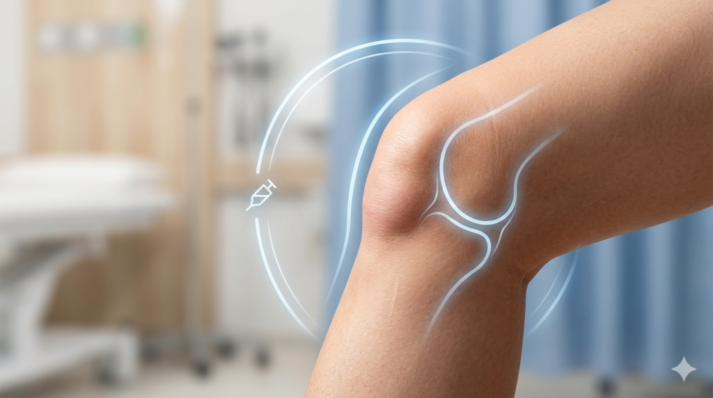
4. Advanced Diagnostics: Arthrocentesis and Fluid Analysis
The definitive step in modern effusion management is arthrocentesis—the sterile aspiration of the joint fluid. This procedure serves a vital dual function:
Immediate Therapeutic Relief: By removing the excess volume, intra-articular pressure is instantly lowered, providing dramatic pain relief and restoring some range of motion.
Precise Diagnostic Analysis: The aspirated fluid is subjected to comprehensive laboratory testing, focusing on:
Viscosity: Inflammatory fluid is thinner (less viscous) than normal or non-inflammatory fluid.
Color/Clarity: Infectious fluid is often cloudy, opaque, or purulent (pus-like); hemorrhagic fluid is bloody.
Cell Count (WBC): Essential for differentiating types of effusion. High counts (>50,000/µL) strongly suggest septic arthritis, while counts between 2,000 and 50,000/µL indicate inflammatory causes (RA, Gout). Counts below 2,000/µL suggest non-inflammatory OA.
Crystal Examination: Polarized light microscopy detects specific crystals for definitive gout or pseudogout diagnosis.
Culture and Gram Stain: Essential for detecting bacterial infections.
5. Management beyond Aspiration: The Modern Treatment Pathway
Traditional treatments relied heavily on resting and systemic NSAIDs. While these remain important, the modern pathway is comprehensive:
Targeted Pharmacotherapy
Treatment now addresses the fluid composition:
For Septic Arthritis: Immediate, targeted intravenous antibiotics.
For Gout: Specific anti-inflammatory agents like Colchicine, and long-term uric acid-lowering therapy.
For RA: Disease-Modifying Antirheumatic Drugs (DMARDs) and biologics designed to arrest the immune attack.
Intra-articular Interventions
Viscosupplementation: In OA, a synthetic hyaluronic acid solution is injected to restore lubricating properties that inflammatory effusion has degraded.
Corticosteroid Injections: Powerful, direct anti-inflammatory action for localized relief.
The Functional Imperative: Physical Therapy
The most significant shift is the emphasis on immediate, controlled movement. Rest (the R in R.I.C.E.) is limited. Physical therapy focuses on strengthening the musculature surrounding the joint (e.g., quadriceps for the knee) immediately after inflammation subsides. This is crucial because a strong muscular system acts as a biological shock absorber, reducing mechanical stress on the synovium and preventing the recurrence of overuse-triggered effusions.
Conclusion: From Passive Symptom to Active Biomarker
Joint effusion must be recognized as a sophisticated biological response, not merely a painful inconvenience. Modern musculoskeletal medicine has transformed how we view and utilize this fluid accumulation. From a systemic warning of gout to a direct metric of osteoarthritis activity through ultrasound tracking, effusion is now recognized as an active diagnostic biomarker. Effective, modern treatment requires moving beyond simple fluid drainage and instead using advanced analysis to unlock precise diagnostic insights and implement targeted, preventative management strategies that preserve joint health for the long term.
FAQ: Frequently Asked Questions in Modern Effusion Management
Q: Can a joint effusion be a sign of a serious, systemic infection? A: Yes. Acute, hot, red, and extremely painful effusion that is not related to a specific trauma must be evaluated immediately for septic arthritis. This is a medical emergency that can lead to permanent joint destruction within 48-72 hours if not treated with antibiotics.
Q: If I have a joint effusion, should I still try to walk/exercise? A: If you suspect an acute traumatic effusion (severe pain, sudden swelling), you must stop and seek medical help immediately to rule out a ligament tear or fracture. For chronic conditions like OA, controlled, low-impact exercise (swimming, cycling) is actually part of the treatment to strengthen the joint, but you should always consult a physical therapist for a personalized plan.
Q: Why would my doctor tell me my joint fluid is “mechanical” vs “inflammatory”? A: This is a key diagnostic distinction that guides treatment. A “mechanical” effusion (low WBC count) suggests the swelling is primarily from mechanical wear (osteoarthritis) and will respond best to rest and physical therapy. An “inflammatory” effusion (high WBC count) means the synovium is actively inflamed (rheumatoid arthritis, gout), requiring specific anti-inflammatory or immunomodulatory medication to arrest the disease process.